Specialized in diagnosing diseases and determining the type of tumor, its malignancy grade, and stage, as well as identifying any tissue markers that affect the tumor's recurrence and aggressiveness. It also involves determining the tumor's response to different types of treatments, whether hormonal, chemotherapy, or targeted therapy, through microscopic examination of tissues and cells.
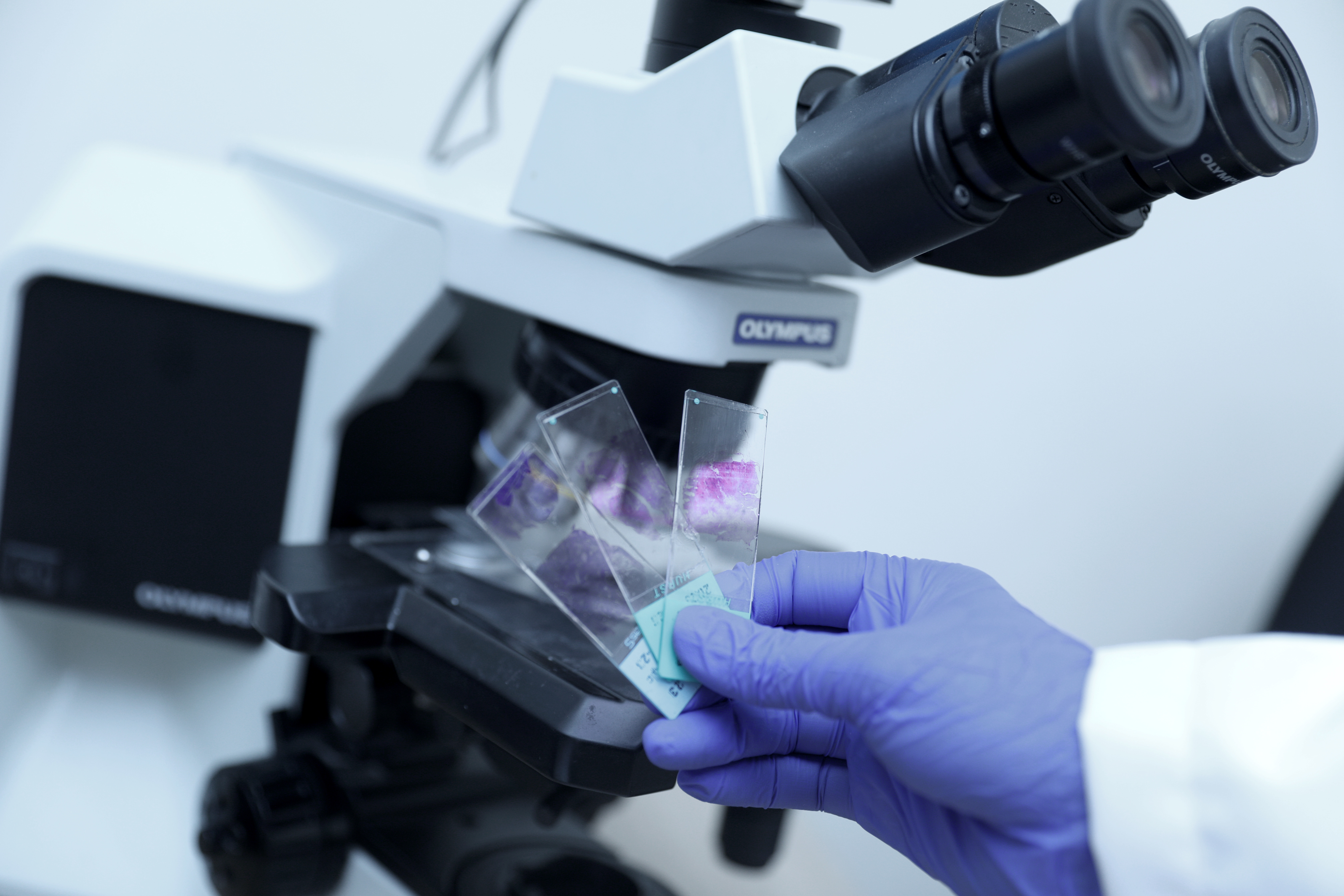
- Pathological examination: This involves examining samples taken by a core needle from the radiology department or surgical operation samples, whether partial or complete excision of the organ or tumor. These samples are received from operations and sectioned for examination.
- Cytological examination: Includes examining fluids from the patient's body and samples from various tissues using a fine needle or examining discharges.
- ROSE clinic (Rapid On-Site Evaluation): These examinations are conducted during the cytological sampling to provide a quick diagnosis, helping the patient reduce the number of samplings needed and ensuring an effective treatment diagnosis. This ensures that an adequate amount of the sample is obtained for all necessary tests, reducing the cost of examination until diagnosis is reached.
- Determining the appropriate treatment: This is done by performing tumor markers and various hormone receptors.
- Frozen section examination: Conducted during the surgical operation to determine the type of tumor and examine the margins of the excised sample to ensure no cancer cells are left in the patient's body after the operation. This is all done while the patient is still in surgery.
- The department retains all parts of the tumor for years to use them in scientific research.
- Doctors: All doctors who examine samples under the microscope, diagnose diseases, and write reports are university faculty members. They hold a Bachelor of Medicine and Surgery, followed by a Master's in Pathology, and a Doctorate in Tumor Pathology for consultants.
We offer a comprehensive range of imaging services
This is the detailed content for Meet the Experts.